Disclaimer:
This article is for information purposes only. It is not a substitute for medical advice or treatment. Seek medical care for your treatment.

What is epilepsy?
Epilepsy is a neurological condition that causes abnormal, frequent seizures. A seizure is a sudden spike in abnormal electrical activity of your brain. When you encounter two or more seizures without any other apparent reason, doctor may diagnose you with epilepsy.
The World Health Organisation (WHO) estimates that 50 million individuals worldwide suffer from epilepsy and the Centers for Disease Control and Prevention (CDC) estimates that approximately 3.5 million individuals in the US have the condition.
Epilepsy can develop in anyone but it is frequent in young children and older adults. According to a research men develop epilepsy more frequently than women. There are two main types of seizures are:
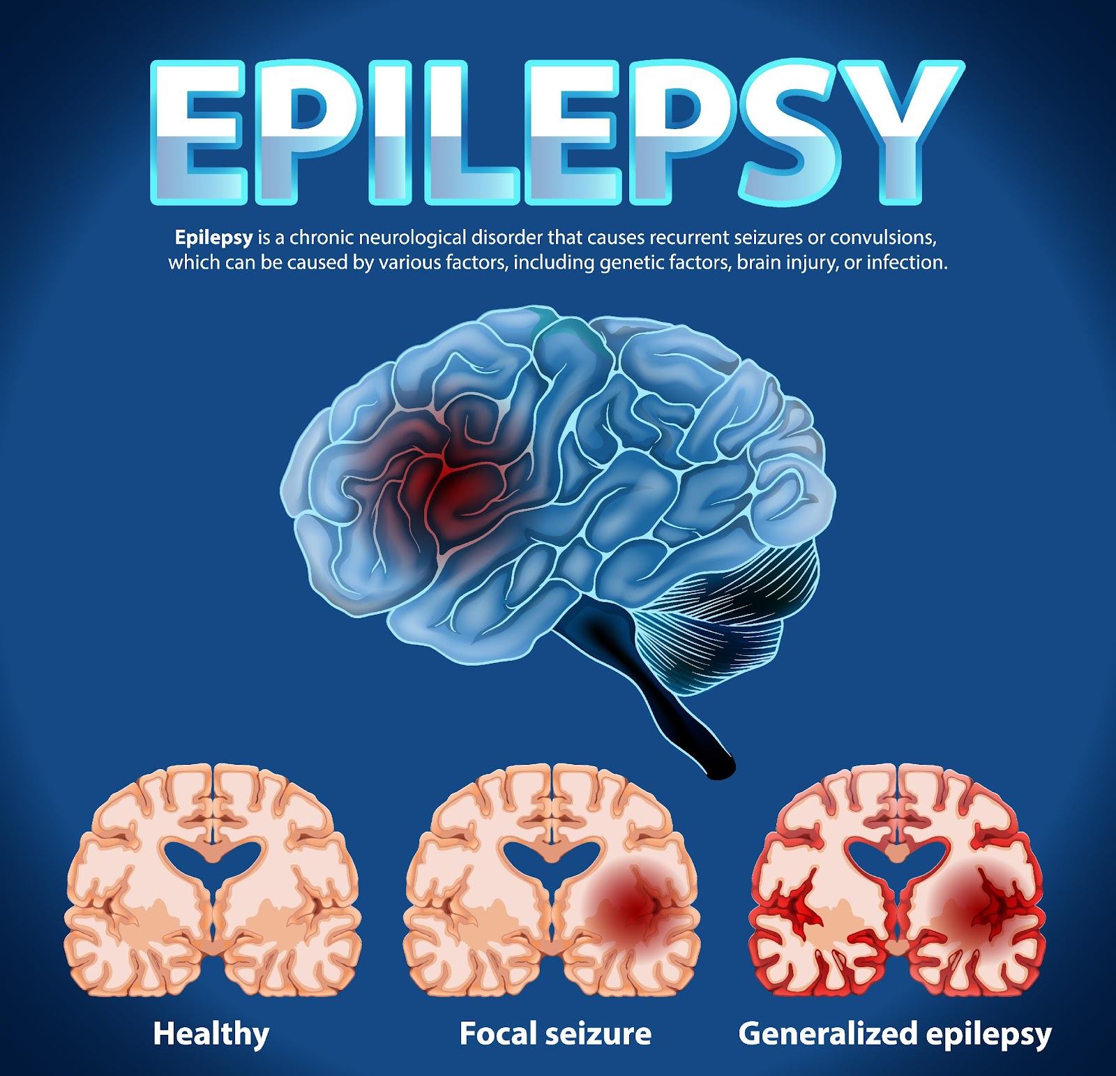
- Generalized seizures: affect your entire brain.
- Focal seizures: affect only a part of your brain.
If you have two or more seizures that were not caused by any known medical condition then you have epilepsy. Your doctor will take a physical exam, take information regarding your medical history, symptoms and order some blood tests before making any diagnosis. Your doctor will ask about certain things during a seizure:
- Muscle jerks
- Muscle rigidness
- Loss of bowel or bladder control
- Irregular breathing
- Pale skin colour
- Blank stare
- Lost consciousness
- Problems in comprehending and talking
Epilepsy diagnosis and tests:
Your doctor may perform several tests to diagnose epilepsy and to identify the cause of seizures. They may include:
- Neurological exam: evaluates your behavior, movements, mental function and other areas. The exam helps in diagnosing epilepsy and identifying the type of epilepsy.
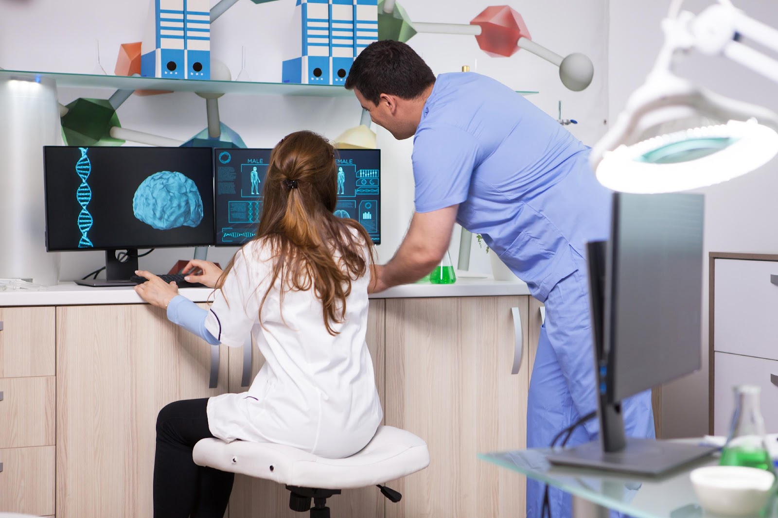
- Genetic testing: leads to more information regarding the condition and treatment of epilepsy in certain people. Genetic testing is generally done in children.
- Blood tests: can identify signs of infections, genetic conditions that may be linked with seizures.
Some brain imaging tests and scans that can diagnose brain changes are:
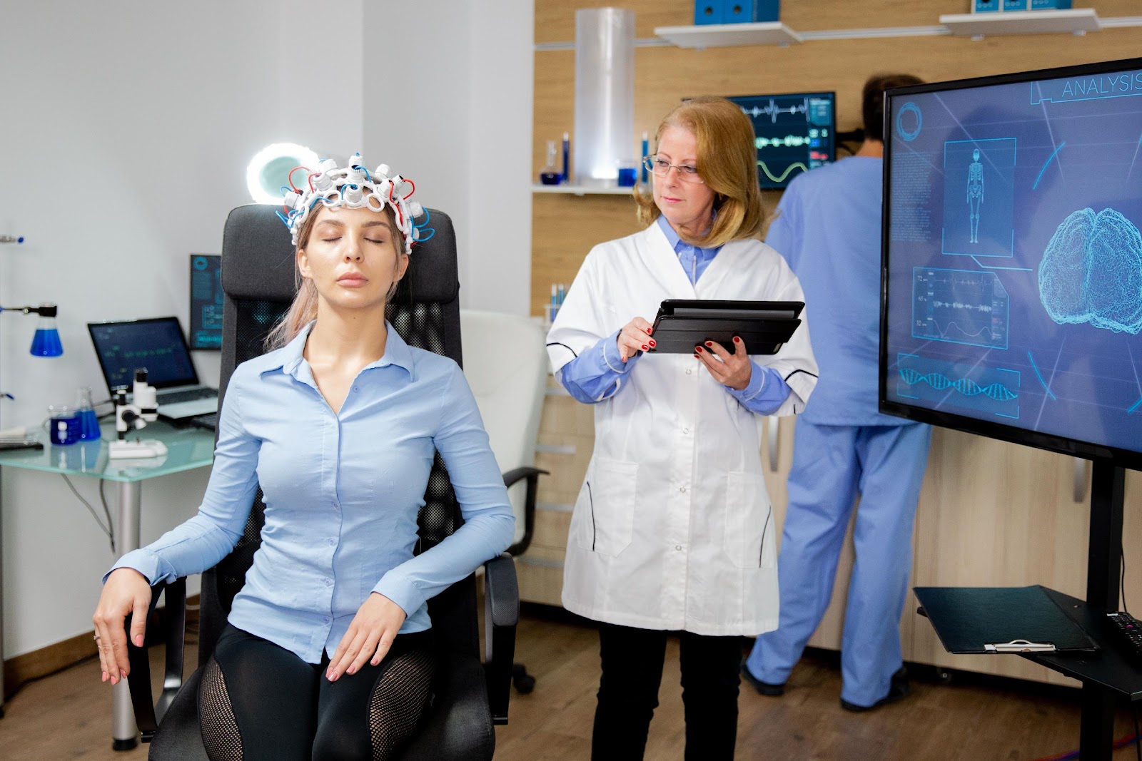
- Electroencephalography (EEG): evaluates the electrical activity in your brain. Some abnormal electrical patterns are linked to the seizures.
- High-density EEG: You might have a high-density EEG in a test that varies from the standard EEG. Electrodes are positioned closer together for this test than they are for a traditional EEG. Your brain’s affected areas may be more specifically identified with a high-density EEG.
- Computerized tomography (CT) scan: A CT scan develops cross-sectional images of your brain using X-rays. CT scans can detect brain cysts, tumors, or bleeding that may be the root causes of epilepsy.
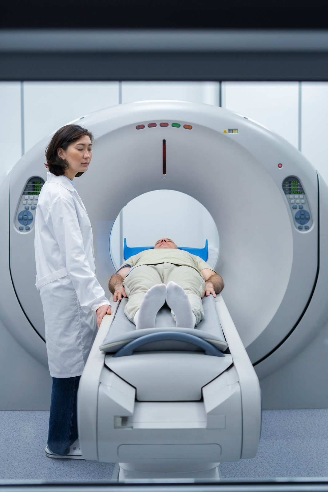
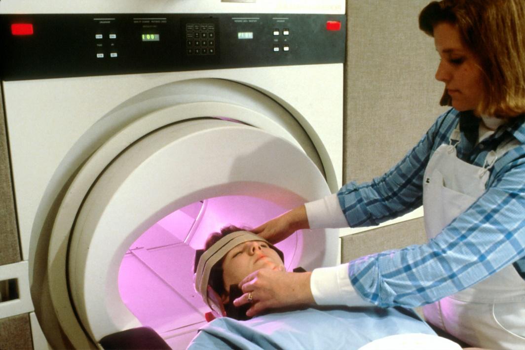
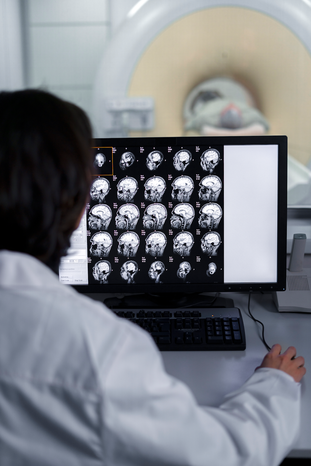
- Magnetic resonance imaging (MRI): Strong magnets and radio waves are employed in an MRI to create a detailed image of the brain. An MRI examines the structure of the brain to identify possible seizure causes, much like a CT scan does. However, a CT scan offers a less detailed view of the brain than an MRI.
- Position emission tomography: scans use little amount of low dose radioactive material. The material is injected into a vein to visualize metabolic activity of the brain and diagnose changes. Parts of the brain with low metabolism may show places where seizure develops.
- Single-photon emission computerized tomography: it is used if MRI and EEG fail to point out the location of the seizures in the brain. SPECT test uses little amount of low dose radioactive material. The material is injected into the vein to create a complete, 3D map of blood flow during seizures.
- Functional MRI: evaluates the changes in blood flow that happen when certain parts of the brain are functional. This test is used prior to surgery to distinguish the right locations of critical functions like speech and movement.
- Neuropsychological tests: these tests evaluate thinking, memory and speech skills. The tests results help identify which areas of the brain are affected by seizures.
Other than tests results a fusion of other techniques may be used to help find out where the seizures begin in the brain:
- Statistical parametric mapping: looks at the areas of the brain with great blood flow during seizures and later compared to the same areas of the brains of people who have no seizures.
- Electrical source imaging: employing an MRI of the brain, EEG data can be shown using the ESI technique. This is done to emphasize the regions that are suffering from seizures.
- Magnetoencephalography: The magnetic fields produced by brain activity are determined by MEG. This helps in identifying probable seizure trigger locations because the brain’s surrounding tissue and skull cause fewer problems with magnetic fields, MEG has the potential to be more precise than EEG. When combined, MEG and MRI can produce images of the brain that display both seizure-affected and seizure-free regions.
Conclusion:
Early diagnosis and treatment can lower seizure frequency and the chances of grave complications. Diagnosing the location and type of your seizure and where it actually begins gives you the ideal chance to discover an effective treatment.